This publication is the edited version of an article that appeared in the September-October issue of the CHR VOICE.
Oct 17, 2025
Dr. David Albertini speaking at the 2019 Activated Egg Symposium in Boston.
Today’s photo gallery features mitochondria, those specialized organelles that we learned in high school and by popular press are the energy-producing powerhouses of all living. Appropriate that we share some imagery regarding mitochondria, given the recent news out of the UK, that 8 babies have now been born using the pioneering technique of pronuclear transfer (see also our next posting on “3-parent pregnancies” on October 20, 2025). The headlines, of course, highlighted this advance as a means for preventing mothers from passing along mutated genes responsible for mitochondrial diseases, a group of quite terrible diseases, often quickly fatal. But such mitochondrial exchange has also been proposed (though by no means confirmed as effective) for older women with infertility.
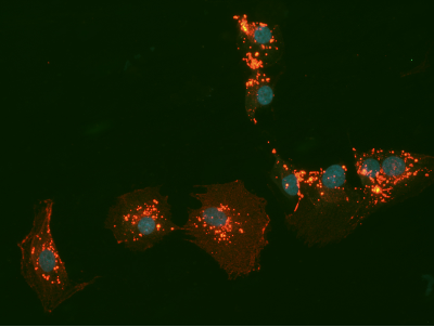
Image 1: The CHR’s researchers have, for years, been interested in the behavior and function of mitochondria in both eggs and granulosa cells. Here is an example of a human granulosa cell cultured and labeled at the CHR, showing in red, the many mitochondria that are distributed throughout the cytoplasm and drive, among other things, the attachment and movement of these cells.
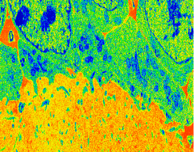
Image 2: This is a pseudo-colored electron micrograph showing the zona pellucida in yellow (lower part) and several cumulus granulosa cells (green, on the top) attached to the zona pellucida. Note the blue structures in the cytoplasm are mitochondria, apposed to the underlying zona, which are believed to provide AROP and other important metabolites to the developing oocyte.
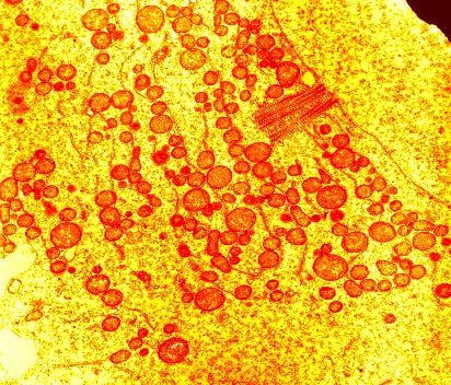
Image 3: This is an electron micrograph of a non-human primate oocyte at an early stage of development. Here, the abundant mitochondria are pseudo-colored red and cluster next to the oocyte nucleus (upper right), forming a structure known as Balbiani’s Body. The internal structure of the oocyte mitochondria suggests that they are inactive and that the oocyte derives its energy resources from the surrounding granulosa cells.
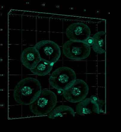
Image 4: This field illustrates a number of actively developing mouse embryos at the transition from two to three cells. Nuclei are visible in each blastomere, and current wisdom suggests that each cell division demands energy resources to advance development, the question being whether these embryos are generating their own fuel from the activity of the mitochondria inherited from the oocyte, or was the gas tank (fuel for development) already filled by granulosa cells during the oocyte’s long journey in the ovary? What is your guess?