Getting a Signature of the Menstrual Cycle — And Profiling Endometrium and Peripheral Blood Cells in Women With and Without Endometriosis
Today’s posting covers two fascinating recent papers focusing on the menstrual cycle. One reported on the plasma proteomic signature of the menstrual cycle, demonstrating that the cycle is accompanied by widespread and notable changes in the circulating proteome. Another addressed the question of how endometrial and systemic immunity is modulated throughout the menstrual cycle. It just goes to show that even when it comes to well-known biological rhythms in the human body, there’s always more to learn.
The CHR’s Editorial Staff

The good old-fashioned menstrual cycle has recently been again in the eye of investigators in two interesting published papers. A first by Danish investigators in Nature Medicine reported the plasma proteomic signature of the human menstrual cycle.1 As the principal motivation for their study, they correctly noted that the menstrual cycle is one of the most fundamental biological rhythms in human physiology. Its systemic molecular changes are, however, still only poorly understood.
They then succeeded in demonstrating that the menstrual cycle is accompanied by widespread changes in the circulating proteome. Specifically, - by profiling nearly 3,000 plasma proteins in 2,760 women from the UK Biobank - they identified 198 proteins that vary across the cycle, forming distinct temporal patterns aligned with menstrual phases, including reproductive hormones, cytokines, and growth factors (and therefore the immune system), many of which are enriched in endometrial tissue and expressed in epithelial as well as stromal cell types. Cycle timing proved to have large effects on only a subset of proteins, while age and BMI explain a larger proportion of variance across a broader set of proteins.
Among many interesting findings, we were especially interested to learn that in pathway enrichment analyses, a cluster of proteins enriched in cytokine signaling, pattern recognition receptor activity, and MAPK pathway regulation – all consistent with immune surveillance, stromal remodeling, and progesterone-driven endometrial maturation following ovulation – was found enriched.
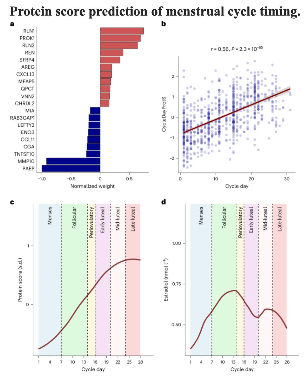
In addition, they were able to link several proteins to common reproductive disorders, including endometriosis, leiomyoma, and just abnormal bleeding, and developed a proteomic score on the basis of 75 proteins that accurately predicts menstrual cycle phase, the latter, of course, having potential relevance to better defining favorable implantation conditions (see the Figure below). On a sidenote, we are on purpose avoiding the term “implantation window.”
More specifically, the study identified 60 significant protein-prevalent disease associations involving 44 unique proteins across 12 conditions. The strongest association was observed between CGA and a history of excessive, frequent, and irregular bleeding. The largest effect estimates were seen for ITIH4 and for IGDCC4 in relation to ovarian cancer.
For incident disease outcomes, the study identified 89 significant protein–disease associations involving cycle day protein comprising 50 unique proteins across eight conditions. The strongest association was observed between CHRDL2 and incident leiomyoma. The largest effect estimate was seen for PAEP and excessive, frequent, and irregular bleeding.
But this is not where this remarkable study stopped; it, next, investigated whether proteins associated with prevalent and/or incident female reproductive diseases play a causal role in disease development or are merely consequences of the disease, using summary statistics for diseases from a recent genome-wide meta-analysis of 42 female reproductive diseases.
Potential causal relationships were identified for 14 protein–disease pairs, among which 13 associations were robust across all sensitivity analyses. Examples included associations between higher SEZ6L2 and risk of endometriosis, higher FSHB and risk of endometriosis, excessive menstrual bleeding, leiomyoma, female genital polyps, and PAEP and risk of excessive menstrual bleeding. Colocalization analyses provided further support to associations between FSHB and endometriosis, excessive menstrual bleeding, and leiomyoma, suggesting a likely shared genetic signal underlying both protein levels and disease risk.
For the proteomic prediction of the menstrual cycle, the study cohort was randomly split into 70% training set (n=1,931) and 30% validation set (n=829). This model then identified 75 proteins with non-zero coefficients. The resulting analysis (cycleDayProts) was strongly correlated with cycle day in the holdout validation set, and its trajectory increased steadily across the follicular phase, reaching a peak in the mid-late luteal phase. As expected, when compared to (known) estradiol levels, they demonstrated a characteristic biphasic pattern, with peaks in the late follicular phase and mid-luteal phase (see below Fig. d) and estradiol correlated weakly with cycleDayProtS). By contrast, cycleDayProtS explained substantially more of the variability in cycle day (R2 = 0.418).
Every published study nowadays includes, in concluding remarks in the discussion section of the paper, a section describing the limitations of the study and, as every study ever published, this study, too, had obvious limitations. But we have to give credit to the authors because only rarely have we seen a limitation discussion as clear and comprehensive as in this paper.
A wonderful paper to read and digest!
And, as noted above, endometriosis was one of the reproductive conditions the Danish study was able to a degree to link to a proteomic signature. And the study, of course, also established a link to cytokines and growth factors linked to the immune system and endometriosis and the immune system have, of course, already for decades been closely linked (and, indeed, the subject of much research at the CHR already several decades ago).
Endometriosis is, indeed, even more closely linked to the immune system than PCOS/PMOS and not only – as widely accepted – as an inflammatory disease but also because endometriosis demonstrates a close statistical association with clinical as well as laboratory evidence of autoimmunity.
This makes a recent paper by investigators from Oxford University in the U.K. – considering that institution’s groundbreaking historical interest in reproductive immunology symbolically housed appropriately in the Peter Medawar Building – relevant to here-discussed menstrual cycle observations when they tried to address the question of how endometrial as well as systemic immunity is modulated throughout the menstrual cycle and whether there exist differences between women with and without endometriosis.2 And the answer they found was that endometriosis was associated with reduced endometrial early natural killer (NK) cells and increased mucosal-associated invariant T (MAIT)-like CD8+ T cells, with cyclical variation.
Though not at the level of the above-discussed Danish study, their results were nevertheless informative. More specifically, compared to controls, patients with endometriosis exhibited decreased endometrial early NK cells and increased MAIT-like CD8+ T cells. The MAIT cells (CD161+Va7.2+CD3+) statistically marginally peaked during ovulation and the implantation window. Peripheral immunity also showed cyclical variation with increased early NK cells and decreased effector CD4 T and effector CD8 T cells in the endometriosis group.
The authors concluded – considering the small number of cases and the marginal statistical significance in the statistical difference, in our opinion, somewhat prematurely – that MAIT cell dysregulation represents a novel feature of endometriosis, potentially contributing to subfertility and providing new avenues for therapeutic development. While that, of course, may ultimately indeed be confirmed, we feel that this study is interesting for two other reasons: (i) It demonstrated locally in endometrium and peripherally rather clear changes in a woman’s immune system over the length of a menstrual cycle; and (ii) the study also again confirmed that local as well as peripheral immune findings in endometriosis patients are different from those of immune systems of control patients.
We found especially the systemically observed findings in blood of interest because blood – in contrast to endometrial tissue – is, of course, always accessible to testing and one, therefore, based on this study, could speculate that expanded data sets could offer new diagnosing potential.
REFERENCES
Riishede et al., Nat Med 2026;32:2311-2318
Kisovar et al., Hum Reprod 2026;deag090
In New York City this September 9th? Come celebrate the Center for Human Reproduction’s 25th Anniversary Gala at the stunning venue Guastavino’s! Reserve your seat here.