News About the Clitoris, Menstrual Effluent, and Thyroid Disorders
Today’s posting covers three interesting news topics related to reproductive medicine and infertility: important discoveries about female sexual anatomy, the potential use of “period blood” for diagnostic purposes, and thyroid function in infertile women. It just goes to show that there is always something new to learn in this field, even though these subjects may not be covered in the mainstream media. As always, we are welcoming comments from our readers.
The CHR’s Editorial Staff

The Clitoris – Likely the Most Under-Studied Organ in Humans – is Suddenly in the News
For centuries, medical science treated female sexual anatomy as secondary or mysterious. As Manuela Callari, a medical writer at Medscape, recently noted, mapping the clitoris — especially its full internal structure and nerve network — has fundamentally changed that view and reshaped women’s health in several important ways.1
First, researchers discovered that the clitoris is not just the small external glans visible at the vulva. Most of the organ is internal, extending deep into the pelvis with erectile tissue, bulbs, and branching nerves surrounding the vaginal canal. Modern imaging and anatomical studies showed it is a complex sensory organ comparable in structural sophistication to the penis.
Older medical models often framed women’s orgasm mainly around vaginal penetration. Clitoral mapping showed that the clitoris is central to female sexual pleasure and orgasm for many women. Researchers now increasingly describe orgasm as involving a broader “clitourethrovaginal complex” rather than a single isolated “G-spot.” This has improved sexual health education, diagnoses of sexual dysfunction, counseling for pain during sex, and treatments in general gynecology. Importantly, precise nerve mapping can have major surgical implications since pelvic surgeries can damage clitoral nerves unintentionally because the anatomy was poorly understood or not even discussed during surgical training.
Recently, precise mapping studies have greatly helped in avoiding such damage during especially vulvar cancer surgeries, reconstructive surgeries, gender-affirming surgeries, and after female genital cutting (in many non-Muslim countries forbidden) or other pelvic trauma. A 2026 report described the first full 3D mapping of the clitoral nerve network, revealing that the nerve branches are more extensive than previously believed. Researchers argued that this could reduce loss of sexual function after surgery.2
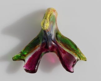
In summary, the clitoris is an intensive internal nerve network, rather than a small external structure. Pelvic surgery can cause long-term sensory loss and impair orgasm, while clitoral mapping may improve episiotomy and vulvar reconstruction in general. Here-noted detailed nerve mapping of the clitoris became possible technically through Synchrotron x-ray imaging by neuroscientist Ju Young Lee, PhD, a research associate at Amsterdam UMC, The Netherlands.
REFERENCES
Callari M. Medscape Medical News June 17, 2026. https://www.medscape.com/viewarticle/mapping-clitoris-reshapes-our-understanding-womens-health-2026a1000kgb
Sims C. The Guardian. March 29, 2026, https://www.theguardian.com/society/2026/mar/29/full-network-clitoral-nerves-mapped-out-first-time-women-pelvic-surgery?utm_source=chatgpt.com
Using Menstrual Effluent for Diagnostic Purposes
Researchers are increasingly treating menstrual effluent (“period blood,” though it also contains endometrial tissue, immune cells, proteins, RNA, lipids, and microbes) as a kind of noninvasive “liquid biopsy” of the uterus. The field is still in its early days, but there are some genuinely promising findings—especially when it comes to endometriosis. A recent systematic review of the subject analyzed 35 studies and found that menstrual effluent consistently reflects known endometriosis biology, including progesterone dynamics, impaired decidualization, inflammatory signaling, immune dysfunction, abnormal angiogenesis, and altered stem cell behavior.1
But there are still important caveats: Studies reported are universally still very small and, therefore, statistically not very solid. Moreover, almost no real-world data exist, - as most studies were performed in highly selected patient populations. Some studies hint at the possibility that patients with infertility and/or recurrent implantation failure (if such a diagnosis really exists) may, indeed, demonstrate abnormal cellular and/or molecular patterns in menstrual effluent.
Menstrual effluent as a research subject is, therefore, attracting increasing attention, as also suggested by a recently published paper in the JCEM, which tried to shed some light on the known association of low vitamin D levels with female infertility.2 The study investigated in healthy females from ages 19 to 40 years the effects of active vitamin D (Calcitrol) on endometrial stromal cell decidualization as defined by menstrual effluent.
Results suggested that vitamin D enhanced decidualization of endometrial stromal cells to a very significant degree (P< 0.0001), - an effect likely produced through reducing protein kinase B (AKT) signaling and further downstream events, suggesting its potential role in female fertility. Vitamin D biomarkers were moreover clearly measurable in menstrual effluent, reemphasizing menstrual effluent as an increasingly interesting research target.
REFERENCES
Watrowski et al., Diagnostics (Basel); 2026;16(5):677. https://doi.org/10.3390/diagnostics16050677
Metz et al., J Clin Endocrinol Metab 2026;11(6):e1532-e1543
What’s the Relevance of Thyroid Disorders for Female Infertility?
Thyroid disorders are highly prevalent during reproductive years, including both thyroid dysfunction and thyroid autoimmunity (TAI). Infertility practice is paying careful attention to thyroid function in infertile women, - though not necessarily because abnormal thyroid function affects female fertility (more on that below), but because even mild hypothyroidism has been associated with lower IQ in offspring.1 Data, however, also suggest that thyroid disease may impair fertility through hormonal and immune-mediated mechanisms.
A recent review article by Belgian colleagues in the JCEM tried to synthesize current evidence regarding effects of thyroid dysfunction, particularly subclinical and overt hypothyroidism, as well as TAI, on female fertility and outcomes of assisted reproductive technology (ART). Especially the role of subclinical hypothyroidism and euthyroid TAI remains controversial.
Interestingly, these investigators in their review article basically ignored the fetal effects of thyroid disease. In concentrating on the effects on female fertility, they concluded that despite limited supporting evidence in the literature, levothyroxine (LT4) is nevertheless widely used in clinical practice, often initiated at thyroid-stimulating hormone (TSH) thresholds of 2.5 mIU/L to define subclinical hypothyroidism and commence treatment.
They then argue that recent data suggest that implementing such thresholds may not be associated with improved fertility outcomes, and that, therefore, higher TSH cutoffs for diagnosis and treatment in the preconception settings may be indicated. They furthermore argued in their paper that ART (i.e., especially IVF) introduces additional considerations, with thyroid status and autoimmunity potentially influencing ovarian response, embryo quality, and pregnancy outcomes and, therefore, concluded that in the light of these uncertainties, treatment decisions increasingly rely on individualized assessment of thyroid function, antibody status, and reproductive context.
Unfortunately – to state it bluntly – even though this review article was accepted in a relatively reputable medical journal, the authors very obviously had no idea what they were talking about. That the JCEM accepted the article and published the paper is moreover clearly a black eye for the mother journal of all Endocrine Society journals. Very obviously, this paper written by medical endocrinologists for a medical endocrinology journal most likely did not see the eyes of a reproductive endocrinologist during the peer review process.
The reasons for this assumption speak for themselves: (i) As already noted above, the threshold of 2.5,IU/L for TSH never had anything to do with a maternal diagnosis of hypothyroidism or with maternal autoimmunity; this cut-off was chosen to not expose offspring to even mild hypothyroidism. This cut-off has absolutely nothing to do with either a diagnosis of hypothyroidism or with consideration of hypothyroidism as a cause of infertility. Indeed, - except for hypothyroidism-associated hyperprolactinemia, - we are unaware of hypothyroidism in any way directly adversely affecting female infertility and/or affecting fertility treatments.
(ii) Similarly, the relationship of autoimmunity is much more complex than the review article reflects. Thyroid disease is, of course, almost always an autoimmune disease. Autoimmune thyroid disease is, indeed, one of the most frequently diagnosed autoimmune diseases during a woman’s reproductive lifespan. But that does not mean that autoimmunity – per se – either through thyroid disease or in other ways – automatically causes infertility.
Though women with thyroid disease trying to conceive are usually, indeed, started on thyroid replacement with thyroxin if the TSH is under 2.5 IU/L, this is done for the benefit of the growing fetus, - and not for the protection of the mother’s well-being. The authors’ argument that there is a growing tendency to - preconceptionally - adopt higher TSH cutoffs for diagnosis and treatment of hypothyroidism by adopting higher TSH, therefore, is non-sensical because the tight control of TSH values is not geared at the mother.
The conceptional mix-up between treating the mother’s potential hypothyroidism to improve her fertility problem – except for correcting potentially her hypothyroid-driven hyperprolactinemia – and preventing a loss of IQ points in the offspring alone, - warrants for this review paper rightful criticism.
REFERENCE
Murphy et al., Austr New Zel Obstet Gynecol 2015;55(5):459-463
Unuane et al., J Clin Endocrinol Metab 2026;111(5):e1239-e1251
In New York City this September 9th? Come celebrate the Center for Human Reproduction’s 25th Anniversary Gala at Guastavino’s! Reserve your seat here.